

Introduction
A muscle injury in the lower back is identified by localized paraspinal tenderness, stiffness, and muscle spasms that worsen with movement but lack radiating nerve pain or leg weakness. To treat a muscle injury in lower back, follow an active recovery protocol: use cold therapy and short walks on days 1 to 2 to calm inflammation; transition to moist heat and gentle range-of-motion stretches on days 3 to 4; and introduce isometric core stabilization exercises (such as bird-dogs and glute bridges) on days 5 to 7 while avoiding prolonged bed rest.
A sudden, sharp catch in the lumbar region can stop you in your tracks. Whether it happens while deadlifting in the gym, reaching into the backseat for a bag of groceries, or simply bending over to tie your shoes, back strain is an almost universal human experience. In the United States, nearly 80% of adults experience at least one debilitating episode of low back pain during their lifetimes. Chronic low back pain affects approximately 13% of adults nationwide, and the combined direct and indirect healthcare burden of back issues reaches an astonishing $365 billion each year.
The good news is that the lumbar spine is remarkably resilient. While severe symptoms can feel terrifying, the vast majority of acute muscle strains begin resolving within days when managed with evidence-based movement strategies rather than prolonged bed rest.
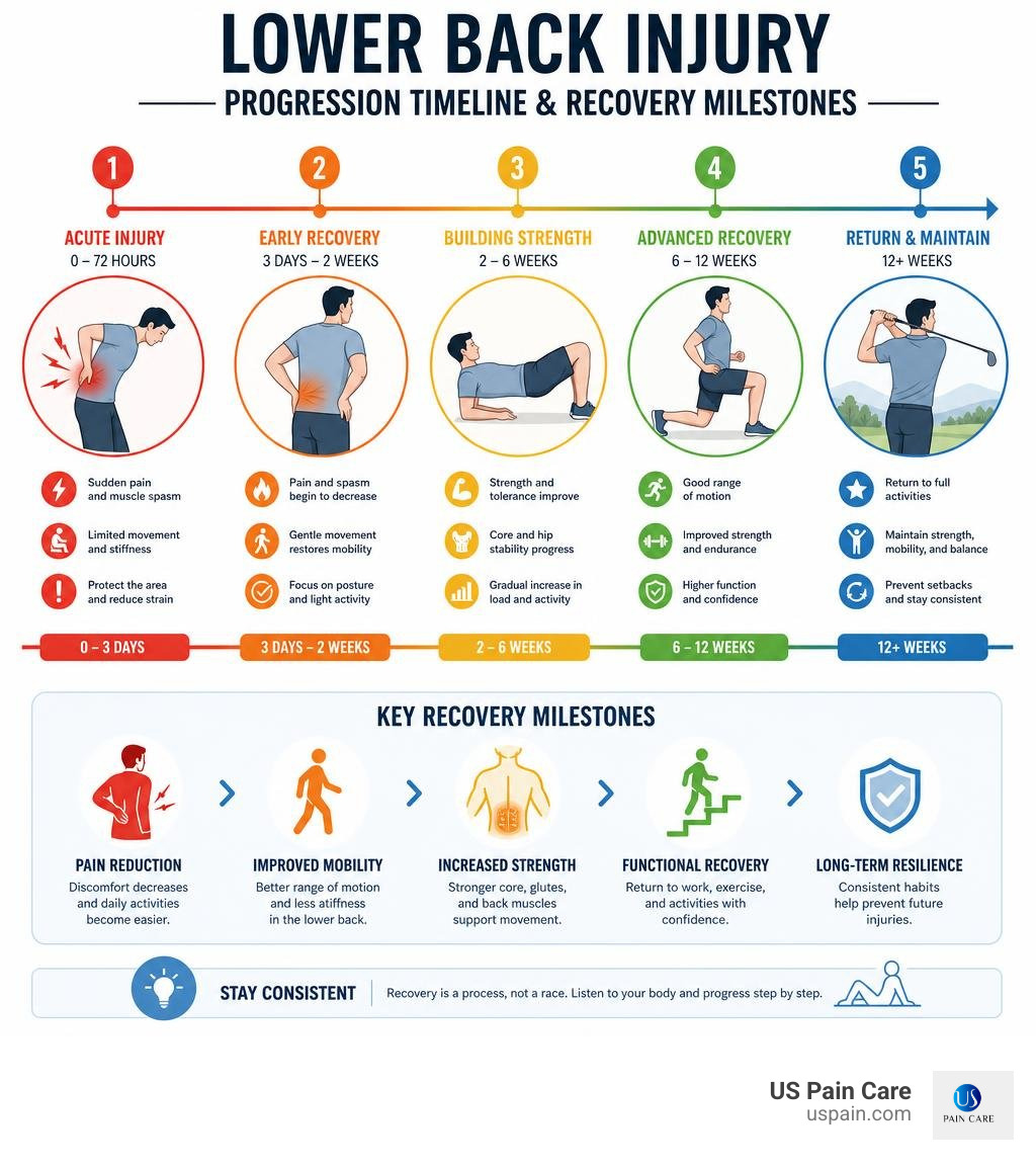
Understanding Muscle Injury in Lower Back: Causes and Anatomy
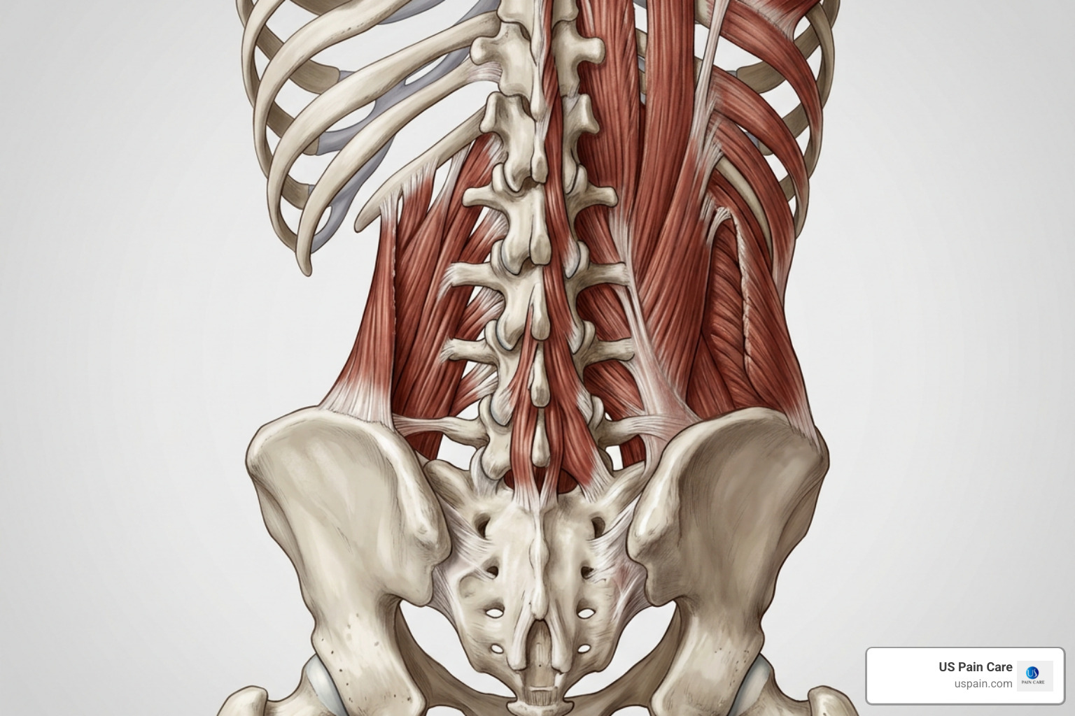
The lower back is engineered to bear the weight of your entire upper body while allowing multi-directional movement. To provide both rigid support and fluid flexibility, the lumbar spine relies on an intricate network of muscles, tendons, and ligaments.
When we experience a muscular strain, microscopic tearing occurs within the muscle fibers or their fibrous attachments. The primary muscular structures involved include:
- Erector Spinae Group: Running vertically alongside the spine (including the iliocostalis, longissimus, and spinalis), these large muscles act as primary spinal extensors, enabling you to stand upright and arch backward.
- Lumbar Multifidus: Deep, segmental stabilizing muscles running along the posterior spine that protect individual vertebral joints from abnormal shearing forces.
- Quadratus Lumborum: A broad, deep muscle connecting the lower ribs to the pelvis, crucial for lateral bending and stabilizing the pelvis during walking.
- Psoas and Core Stabilizers: Anterior and lateral muscle groups that work in tandem with posterior muscles to create intra-abdominal pressure.
According to clinical research on mechanical back strain (Yusupov et al., 2026), lumbar strains account for nearly 70% of all mechanical low back pain episodes. About one-third of individuals cannot pinpoint a single dramatic event; rather, the injury develops from cumulative micro-trauma.
Common culprits include violent muscular contractions during lifting, sudden awkward twisting, prolonged sitting with poor pelvic tilt, tight hamstrings that tug on the pelvis, or sudden axial overload. For those looking for a rapid breakdown of early symptoms, our A Quick Start Guide to Back Muscle Strain provides a helpful introductory framework.
Microscopic Changes and Muscle Degeneration
When lower back tissues are strained, the damage extends beyond temporary surface soreness. At the cellular level, acute trauma triggers an inflammatory cascade. If underlying spinal instability or persistent nerve irritation is present, deeper postural muscles can undergo long-term microscopic remodeling.
Recent investigations into spinal pathology—such as a landmark study on multifidus pathology—have revealed that structural abnormalities in muscle tissue correlate closely with ongoing pain and functional disability. Researchers observed distinct “moth-eaten,” split, and lobulated muscle fibers alongside Type I (endurance) fiber atrophy in affected lumbar segments.
Furthermore, a comparative study on lumbar muscle degeneration (Öztürk & Aylanç, 2020) demonstrated that chronic back issues often involve significant fatty infiltration and degeneration within the erector spinae and multifidus muscles, particularly in individuals over age 40 and women. When deep stabilizers lose their structural integrity, the spine becomes increasingly prone to recurring injury, highlighting why active rehabilitation is critical.
Distinguishing Muscle Strains from Serious Lumbar Conditions
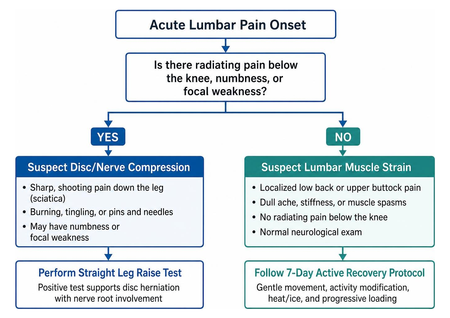
Differentiating a benign muscular issue from discogenic or neurological conditions is essential for safe recovery. Muscular strains typically present with:
- Localized tenderness to deep palpation across the paraspinal muscles.
- Pain that worsens with active movement (bending, twisting, standing up) and eases when lying supported.
- Visible or palpable muscle spasms.
- Stiffness and reduced range of motion without neurological deficits.
If you are experiencing severe cramping or spasms, exploring Lower Back Pain and Spasms Treatment options can help you calm hyperactive muscle guarding.
| Clinical Feature | Lumbar Muscle Strain | Lumbar Disc Herniation | Lumbar Spinal Stenosis |
|---|---|---|---|
| Primary Pain Location | Localized to lumbar paraspinals, beltline, or upper buttock | Low back with sharp, shooting pain down the leg (radiculopathy) | Bilateral buttocks, thighs, and calves |
| Sensation | Dull ache, stiffness, localized soreness, muscular spasms | Burning, electric-shock sensation, tingling, pins and needles | Aching, heaviness, cramping, numbness in legs |
| Aggravating Factors | Bending, twisting, lifting, prolonged standing | Sitting, coughing, sneezing, forward flexion | Walking downhill, prolonged standing upright |
| Relieving Factors | Gentle walking, lying supine with knees bent, heat/ice | Standing, walking, lumbar extension | Sitting down, leaning forward (“shopping cart sign”) |
| Neurological Exam | Normal reflexes, motor strength, and sensation | Often positive straight leg raise, focal weakness, reflex deficit | Generalized sensory changes, balance deficits |
Differentiating a Muscle Injury in Lower Back from a Herniated Disc
A classic muscle strain stays relatively contained within the lumbar and gluteal zones. In contrast, lumbar disc herniation involves displacement of the intervertebral disc’s inner nucleus pulposus through a torn outer annulus fibrosus, which frequently compresses adjacent spinal nerve roots.
According to evidence-based guidelines on lumbar disc herniation (Qaraghli & Jesus, 2023), disc herniations occur at an annual rate of 5 to 20 cases per 1,000 adults, most commonly at L4-L5 or L5-S1. A herniated disc typically produces true radicular pain (sciatica)—sharp, burning pain radiating below the knee into the calf or foot, accompanied by dermatomal numbness or motor weakness (such as foot drop). Clinicians evaluate this using the Straight Leg Raise (Lasègue) test.
To dive deeper into the nuances of localized soft tissue strain versus disc pathology, see our Detailed Guide to Relieving Lower Back Muscle Sprain.
Diagnostic Protocols and Imaging Criteria
For acute, uncomplicated lower back pain without red flags, routine imaging (X-rays, CT scans, or MRIs) is not recommended during the initial 4 to 6 weeks. Scans performed prematurely often identify incidental findings—such as age-appropriate disc bulges or facet arthrosis—that do not correlate with the patient’s acute symptoms and may cause unnecessary anxiety.
A comprehensive clinical physical examination is the gold standard for initial assessment. Imaging is strictly indicated if conservative care fails after 6 weeks or if red-flag symptoms appear:
- Loss of bowel or bladder control (incontinence or urinary retention).
- “Saddle anesthesia” (loss of sensation in the perineum, groin, or buttocks).
- Rapidly progressing neurological weakness in the legs.
- Unexplained weight loss, history of cancer, severe fever, or chills.
- High-velocity trauma (e.g., motor vehicle collision, serious fall).
Step-by-Step Recovery: 7-Day Protocol for Acute Pain Relief
When treating an acute strain, the historic advice of “strict bed rest for a week” has been completely disproven. Prolonged immobility causes rapid deconditioning, stiffens paraspinal tissues, and can prolong recovery. Modern rehabilitation emphasizes active rest—modifying painful activities while maintaining gentle, regular movement.
To navigate this process safely, follow our insights on Healing Your Pulled Lower Back Muscle the Right Way.
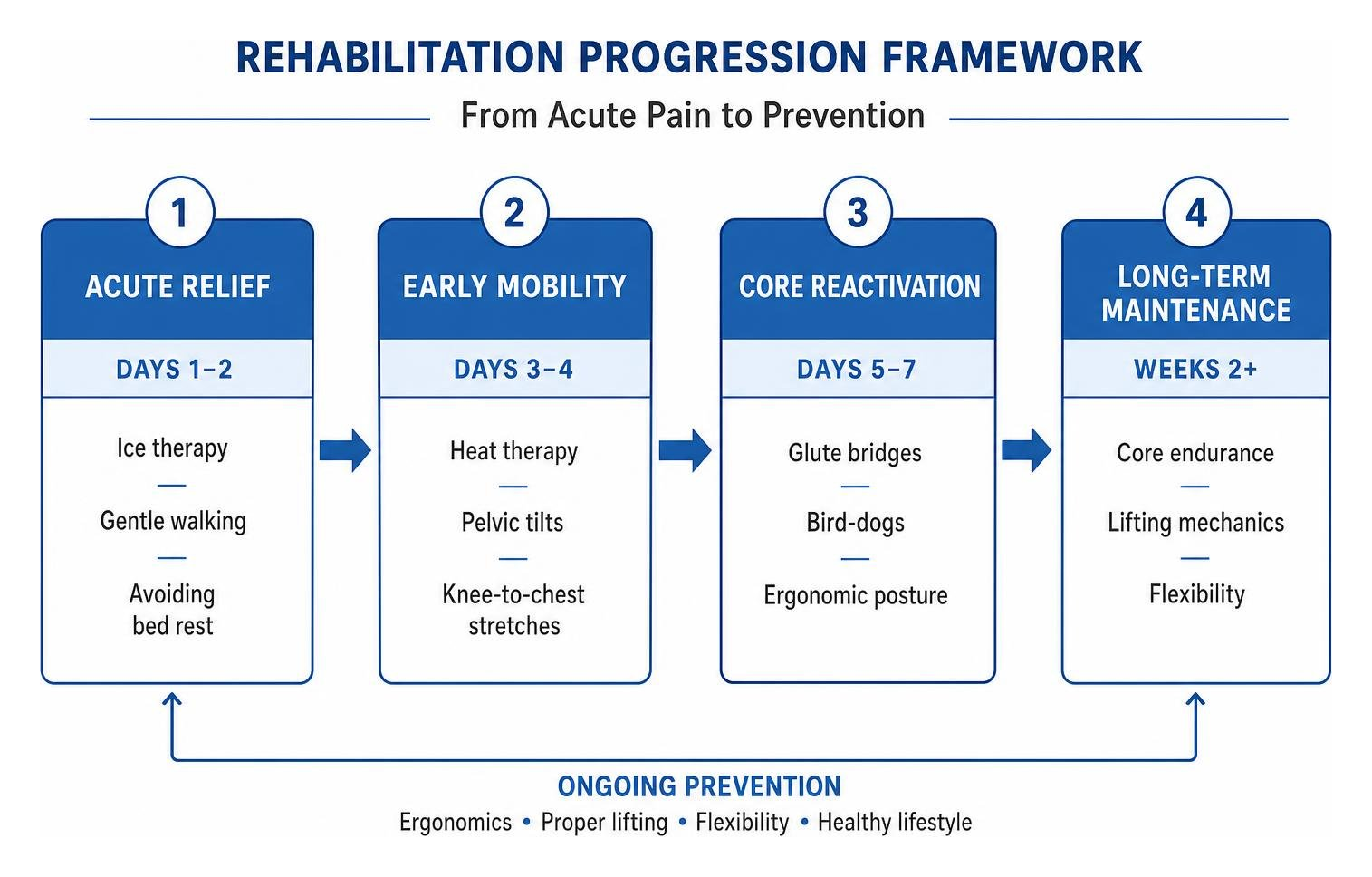
7-Day Action Plan for Muscle Injury in Lower Back Recovery
Days 1 to 2: Calm the Acute Spasm and Reduce Inflammation
- Cryotherapy: Apply ice packs wrapped in a thin towel for 15 to 20 minutes every 2 to 3 hours. Cold therapy numbs irritated nerve endings and curbs local inflammatory edema.
- Short, Frequent Walks: Avoid prolonged sitting or lying down. Walk on a level surface for 5 to 10 minutes every few hours.
- Supported Resting Positions: Lie on your back with a pillow beneath your knees, or lie on your side with a pillow between your knees to take mechanical tension off the lumbar spine.
- Over-the-Counter Analgesia: Non-steroidal anti-inflammatory drugs (NSAIDs like ibuprofen or naproxen) or acetaminophen can provide short-term symptomatic relief if medically appropriate.
Days 3 to 4: Introduce Gentle Heat and Tissue Mobility
- Transition to Heat: Once the sharpest pain subsides (typically after 48 hours), apply moist heat packs for 15 to 20 minutes to improve local circulation, soothe hypertonic muscles, and reduce stiffness.
- Gentle Range-of-Motion Exercises:
- Pelvic Tilts: Lie on your back with knees bent. Gently flatten your lower back against the floor by tightening your abdominal muscles, hold for 3 seconds, and release. Repeat 10 times.
- Knee-to-Chest Stretches: While supine, slowly draw one knee toward your chest until a comfortable, pain-free stretch is felt in the lower back and glute. Hold for 15 seconds; alternate sides.
- Maintain Regular Walking: Gradually increase walking sessions to 15 minutes twice daily.
Days 5 to 7: Reactivate Core Stabilizers and Restore Function
- Isometric Core Engagement:
- Bird-Dog: On hands and knees, slowly extend one arm forward and the opposite leg straight back while keeping the spine neutral. Hold for 3 seconds, return, and alternate.
- Glute Bridges: Lie supine with knees bent and feet flat. Squeeze your glutes and lift your hips until your thighs and torso form a straight line. Hold for 3 seconds, lower slowly, and repeat for 2 sets of 8 to 10 repetitions.
- Postural Awareness: Avoid slouching in soft couches or chairs. Use a small lumbar roll when sitting, and take standing micro-breaks every 30 minutes.
For detailed demonstrations and technique instructions, review How to Perform Safe Recovery Exercises for Low Back Strain.
Long-Term Rehabilitation and Recurrence Prevention
While more than 90% of individuals recover from an acute episode of lumbar strain within 4 weeks, long-term recurrence rates are notably high. Between 35% and 69% of patients experience another episode within 12 months, with a median time to recurrence of about 139 days.
Preventing a relapse requires addressing the underlying biomechanical deficiencies that contributed to the initial strain:
- Progressive Core and Lumbar Stabilization: Transition from basic home stretches to structured strength training. Building endurance in the transverse abdominis, multifidus, and gluteal musculature provides a protective muscular corset around the lumbar vertebrae.
- Proper Ergonomics and Workstation Setup: Adjust office chairs to support the natural inward curve of the lumbar spine. Keep computer monitors at eye level, position feet flat on the floor, and avoid sitting uninterrupted for hours.
- Ergonomic Lifting Mechanics: Always lift from the legs by bending at the hips and knees—never bend from the waist with straight legs. Keep heavy objects close to your body’s center of gravity and avoid twisting your torso under load.
- Lower-Body Flexibility: Maintain flexibility in the hamstrings, hip flexors, and piriformis muscles to preserve normal pelvic alignment and prevent excessive strain on the lower back.
- Lifestyle Factors: Quitting smoking or vaping improves microvascular blood flow to spinal tissues, and managing body weight decreases chronic axial load on the lumbar spine.
For individuals facing persistent or complex symptoms, integrating our step-by-step multidisciplinary back strain management approach helps address the physical, postural, and behavioral aspects of spinal recovery.
Frequently Asked Questions about Lower Back Strains
How long does it take for a strained lower back muscle to heal?
The vast majority of acute lumbar muscle strains resolve within 10 to 14 days, with more than 90% of patients achieving complete functional recovery within one month. Complete structural tissue remodeling can take up to 6 weeks. Healing times vary based on overall physical conditioning, smoking status, and adherence to early, gentle mobilization.
Should I rest in bed or keep moving after pulling a back muscle?
You should stay as active as your pain allows and avoid strict bed rest. Clinical research confirms that prolonged bed rest (more than 24 to 48 hours) leads to muscle deconditioning, joint stiffness, and increased pain duration. Light daily activities and short, regular walks promote blood flow and accelerate tissue healing.
When is lower back muscle pain considered an emergency?
Seek emergency medical evaluation immediately if your back pain is accompanied by red-flag symptoms: sudden loss of bowel or bladder control, numbness in the saddle/groin area, progressive weakness in your legs or feet (such as foot drop), high fever, or if the pain follows major physical trauma.
Conclusion
Experiencing a muscle injury in the lower back can disrupt your daily routine, but it rarely signals permanent damage. By following an active 7-day protocol—combining cold and heat therapy, short walks, gentle mobility drills, and progressive core stabilization—you can safely overcome acute discomfort and protect your spine from future flare-ups.
At US Pain Care, our board-certified pain management physicians specialize in whole-person, multidisciplinary care. From advanced diagnostics and targeted physical therapy to cutting-edge, minimally invasive interventions, we help patients overcome stubborn pain and return to the activities they love.
If your back pain persists beyond a week, worsens with movement, or limits your ability to live comfortably, consider seeking clinical evaluation when you pulled something in your lower back to receive an individualized, physician-led treatment plan.
References
David Yusupov, Minh Quan T. Le, Todd May. “Mechanical Back Strain.” StatPearls Publishing, 2026. NBK542314. Öztürk PE, Aylanç N. “Which is the most affected muscle in lumbar back pain – multifidus or erector spinae?.” Polish journal of radiology, 2020. PMCID PMC7361368. Mustafa I. Al Qaraghli, Orlando De Jesus. “Lumbar Disc Herniation.” StatPearls Publishing, 2023. NBK560878.